Realistic Simulations 3D

Datasets of the Challenge 2016
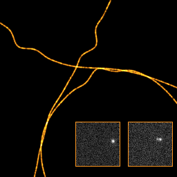
The benchmarking of SMLM software package mainly relies on the usage of common reference datasets. This page contains a collection of localization microscopy simulated datasets that were used to conduct the challenge 2016. They are provided in 2D and in various 3D modalities AS astigmatism, BP biplane, and DH double-helix.
Reference: Sage et al., Super-resolution fight club: assessment of 2D and 3D SMLM software Open-access PDF, Nature Methods 2019.
| Datasets | Simulated structure | Noise level | Density | Modality | Number of frames | Ground-truth |
|---|---|---|---|---|---|---|
| Datasets with ground-truth localizations (publicly available) | ||||||
| MT0.N1.LD | Microtubules | N1 High SNR | LD Low density (0.2) | 2D, AS, BP, DH | 19'996 | Yes |
| MT0.N1.HD | Microtubules | N1 High SNR | HD High density (2) | 2D, AS, BP, DH | 2'500 | Yes |
| MT0.N2.LD | Microtubules | N2 Low SNR | LD Low Density (0.2) | 2D, AS, BP, DH | 19'996 | Yes |
| MT0.N2.LD | Microtubules | N2 Low SNR | HD High density (2) | 2D, AS, BP, DH | 2'500 | Yes |
| Contest datasets with hidden localizations | ||||||
| MT1.N1.LD | Microtubules | N1 High SNR | LD Low Density (0.2) | AS, BP, DH | 19'996 | No |
| MT2.N1.HD | Microtubules | N1 High SNR | HD High density (2) | AS, BP, DH | 3'125 | No |
| MT3.N2.LD | Microtubules | N2 Low SNR | LD Low Density (0.2) | AS, BP, DH | 20'000 | No |
| MT4.N2.HD | Microtubules | N2 Low SNR | HD High density (2) | AS, BP, DH | 3'020 | No |
| ER1.N3.LD | Endoplasmic Reticulum | N3 Very low SNR | LD Low Density (0.2) | 2D | 19'620 | No |
| ER2.N3.HD | Endoplasmic Reticulum | N3 Very low SNR | HD High density (5) | 2D | 3'020 | No |
| Calibration | ||||||
| Beads | Z-Stack of beads | High SNR | 6 beads / slice | 2D, AS, BP, DH | 151 slices-750nm to 750nm | No |
Note of noise levels
- N1: Typical photon counts and background levels for Alexa647 labelled STORM sample.
- N2: Photoswitchable fluorescent protein labelled sample such as mEos2 or Dendra2.
- N3: Typical photon counts and level of background for site specific dye-labelled live cell STORM sample such as ER Tracker.
Synthesis of the realistic datasets
The synthetic datasets were designed to be as similar as possible to images derived from cellular structures in real experimental conditions To achieve the high degree of realism, we defined mathematical models for biological structure that try to imitate microtubules and endoplasmic reticulum/mitochondria. These structure have a tubular shape in the 3D space. Typically, microtubules are defined with their central axis elongating in a 3D space having an average outer diameter of 25 nm with an inner, hollow tube of 15 nm diameter.
The underlying sample structure is formalized in a continuous space which allows rendering of digital images at any scale, from very high resolution (up to 1 nm/pixel) to low resolution (camera resolution: 100 nm).The continuous-domain 3D curve is represented by means of a polynomial spline. The sample is imaged in a limited field of view, i.e., less than 6.4 × 6.4 μm2, and the center lines of the microtubules have limited variation along the z (vertical) axis, i.e., less than 1.5 μm. The fluorescent markers are uniformly distributed over the structure according to the required density. The photon emission rate of each fluorophore is controlled by a photo-activation model (see below).
The exact locations of all fluorophores are therefore stored at high precision, as floating point numbers expressed in nanometers. This ground-truth file is useful for conducting objective evaluations without human bias.