Results Tubulin-A647-3D
3D astigmatic dSTORM image of nuclear pore complex. Courtesy of Jonas Ries (EMBL)
Sage et al., Quantitative Evaluation of Software Packages for Single-Molecule Localization Microscopy Open-access PDF, Nature Methods 2019. Li et al., Real-time 3D Single-Molecule Localization ising Experimetal PointSpread Functions Open-access PDF, Nature Methods 2018.

Convention reconstruction
Integrating all the frames
Tubulin-A647-3D • 2D
Real dataset Courtesy of Jonas Ries (EMBL)
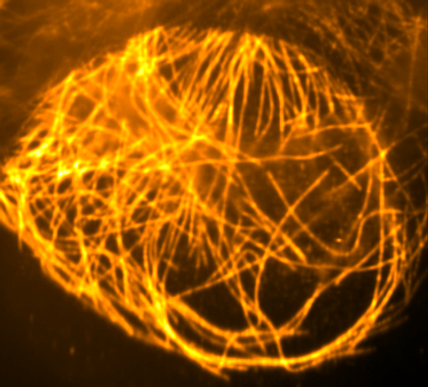
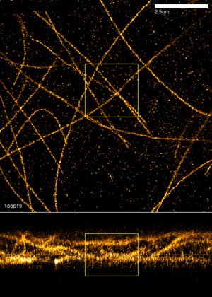
- 3D astigmatic image of microtubules imaged using dSTORM with a cylindrical lens (112'683 frames)
- Microtubules in U-2 OS cells, labeled with anti-alpha tubulin primary and Alexa Fluor 647-coupled secondary antibodies.
- Calibration data for the tubulin dataset. Fluorescent beads adsorbed to a glass coverslide in water are stepped in Z at high SNR
Download Dataset Tubulin-A647-3D
| File | Link to Download | Size |
| Tubulin-A647-3D-stacks_2.tif | Tubulin-A647-3D-stacks_2.tif | 0 Mb |
| ThunderSTORM-Tubulin_30nmpix.tif | ThunderSTORM-Tubulin_30nmpix.tif | 0 Mb |
| Tubulin-A647-3D-stacks_4.tif | Tubulin-A647-3D-stacks_4.tif | 0 Mb |
| Tubulin-A647-3D-stacks_3.tif | Tubulin-A647-3D-stacks_3.tif | 0 Mb |
| Tubulin-A647-3D-stacks_1.tif | Tubulin-A647-3D-stacks_1.tif | 0 Mb |
| Tubulin-A647-3D-BEADS-as-sequence.zip | Tubulin-A647-3D-BEADS-as-sequence.zip | 0 Mb |
| Tubulin-A647-3D-BEADS-as-stacks.tif | Tubulin-A647-3D-BEADS-as-stacks.tif | 0 Mb |
